


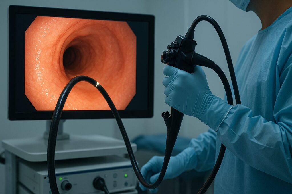
A prospective, single-center study published in the Journal of Clinical Medicine evaluated AI-assisted lesion detection and disease-specific optical classification during surveillance colonoscopy in patients with ulcerative colitis (UC). The study included 82 adults undergoing three sequential colonoscope withdrawals using high-definition white-light imaging (WLI), linked-colour imaging (LCI), and LCI with Fujifilm’s CAD-EYE AI detection system activated. Among 54 patients with identified lesions, researchers found 281 lesions, including 22 neoplastic lesions in 19 patients. Most neoplastic lesions were low-grade dysplasia; one was high-grade dysplasia, and no adenocarcinomas were identified.
The overall lesion miss rate was 5.7% with WLI, 2.5% with LCI and 0% with CAD-EYE-assisted examination. The differences between CAD-EYE and both other methods were statistically significant. However, differences in neoplasia miss rates (18.2%, 4.5% and 0%, respectively) were not statistically significant. For lesion characterization, the disease-specific Kudo-IBD classification achieved the highest diagnostic accuracy at 85.8%, compared with 74% for CAD-EYE, 74% for conventional Kudo and 64.4% for NICE. Kudo-IBD also demonstrated 90.9% sensitivity, 85.3% specificity and a 99.1% negative predictive value.