Key Points:
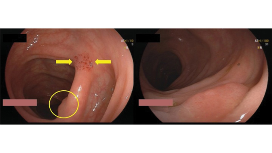
- Flat lesions are commonly found during colonoscopies but can be difficult to keep track of, especially in less-than-ideal conditions such as an unclean colon, increased colon motility, or when the scope moves away from the lesion.
- The technique involves focusing on the lesion, pushing the scope towards it, and then suctioning the mucosa next to it. This action creates a red submucosal suction mark, which serves as a pivotal reference point to re-identify or relocate the polyp.
- The technique consists of three main steps: application, reshape, and resection.
- Application: The flat lesion is located and the scope is placed directly on it. Suction is then applied to draw the lesion into the working channel.
- Revisualization and Reshape: Suctioning creates a mark and reshapes the polyp into a pseudo-polyp or “sessile” lesion. During suction, a snare is inserted into the working channel, transforming the flat lesion into a more visible and marked sessile lesion.
- Execution (Resection): Advancing the snare displaces air in the working channel and likely detaches the suctioned polyp. Detachment also occurs when suction is discontinued. If the scope position is lost, the marked polyp is easier to find. If the position is maintained, immediate resection (cold or hot) is performed.


